Alaska Blind Child Discovery
A
cooperative, charitable research project to vision screen every preschool
Alaskan
Nasolacrimal Duct Obstruction
Home
Tears are an important part of the health and function of the eye. A smooth tear film is the most important optical surface of the eye. The continuous rinse of tears with their immunoglobulin-A provides an excellent barrier to infection and irritants. Without adequate tears, the eye can be blurry, infected, scarred and, eventually blind. A major cause of world blindness is dry eye!
Our tears are composed to three layers for optimum health. Mucous glands in the conjunctiva produce mucous adjacent to the eye surface. Oil glands in the lids produce an oil layer on the outside to prevent evaporation. Sandwiched between the mucous and oil is the salt-water layer normally produced by multiple small glands. Pain, cold wind and emotions can cause an outpouring of extra salt water from the large gland under the brow. Some infants do not well-up with tears even when crying; their large glands are not yet fully functional but the eyes are safely protected by multiple small glands.
Tears require active blinking to cover the eye. The eye must shut completely during sleep to avoid areas of drying. Eventually tears leave the eye through the nasolacrimal duct system (Figure 1). Tears find their way to the nose and eventually are swallowed.
Causes of tearing
Excess tearing in children is most commonly caused by a blocked tear duct. Also known as nasolacrimal duct (NLD) obstruction, the tearing is associated with a white eye and discharge. Excess tearing is also a symptom of several other serious eye diseases. Painless tearing with matter (mucopurulence) can also be caused by bacterial or viral conjunctivitis ("Pink Eye"). Infections, foreign bodies or damage to the cornea (keratitis) will cause a painful, red eye and excess tearing. Tearing can be caused by abnormal lid shape or misaligned eyelashes rubbing the eye. Inflammation on the inside of the eye (uveitis) will cause a painful red eye with tearing. The hallmark of vision-threatening congenital glaucoma (high eye pressure) is a steamy cornea with excess clear tearing. Abnormal rearrangement of nerves in the face can increase tearing or cause tears while eating (Crockodile Tears). Inadequate blinking may result in excess tearing. Excess tearing may also be caused by visual disturbances causing blur or double vision.
Anatomy
Figure 1. The Anatomy of the Tear System.
At the nasal side of each eyelid is a tiny opening (punctum) leading to a narrow canaliculus (Figure 1). Canaliculi from upper and lower lids combine and empty into the nasolacrimal sac. The Valve of Hasner separates the bottom of the sac from the nose. Nasolacrimal Duct Obstruction is due to a membrane or bony blockage of the Valve of Hasner. There are also Rosenmüller valves in the canaliculi which prevent tears and air from flowing back toward the eye. If all three valves are blocked at birth, a blue cyst forms at 1-2 days of life requiring urgent treatment to prevent serious infection (Dacryocele).
Frequency and Natural Course of NLD Obstruction
More than 6% of newborns have nasolacrimal duct obstruction in at least one side. NLD obstruction is more common in children with frequent ear infections. With conservative observation, and occasional antibiotic drops, almost all cases of NLD obstruction clear by age 7 months or 18 pounds (Figure 2). Spontaneous resolution occurs because most obstructions are due to a membrane at the Valve of Hasner which stretches and opens with facial bone growth. Rarer NLD obstructions are caused by tight bones blocking the Valve of Hasner.
Figure 2. The Natural history of congenital nasolacrimal duct obstruction.
Figure 3. Left nasolacrimal duct obstruction caused by bony obstruction at the Valve of Hasner.
These will persist with episodes of excess tearing and discharge for years. Occasionally the tear sac infections burst into surrounding tissues adjacent to the eyeball causing a life-threatening infection (Orbital Cellulitis). Gentle massage of the nasal side of the lids in NLD obstruction is recommended by many pediatricians. This technique helps mix antibiotic drops with the infections in the tear sac. Be careful to massage the tear sac GENTLY, since overzealous massage can produce orbital cellulitis.
Treatment
Persistent NLD obstructions and dacryoceles are treated by gently passing a flexible, metal probe through the punctum, canaliculus, sac and Valve of Hasner. In newborns, this can be done awake with the head held firmly to avoid accidental trauma to the canaliculus. Larger infants usually require a brief anesthetic; intubation prevents aspiration of pus and blood. In case of NLD obstruction probed later than 8 months, approximately 15% re-obstruct. To avoid this recurrance, a silicone stent can be passed and left in place so the Valve of Hasner can heal open (Figure 4).
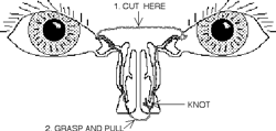
Figure 4. Silicone stent intubation as treatment for nasolacrimal duct obstuction. The "Farson" modification allows easy home removal since each side is treated with one silicone loop.The external portion of the stent is cut and pulled for removal after one month as an an outpatient.
Dr. Clyde Farson of the Alaska Native medical Center suggested a modification of silicone intubation for NLD obstruction (both sides with one silicone tube stent) which is easily removed at home by parents. The "Farson" modification saves time and money especially for families living long distances from their eye doctor.
Conclusion
Most children see well because they have adequate, protective tears. Inadequate amount or distribution of tears can cause serious illness. Excess tearing can also indicate serious eye disease. Fortunately, excess tearing in babies is usually due to nasolacrimal duct obstruction, most cases of which spontaneously resolve in 7 months. Atypical or persitent tearing symptoms should be brought to the attention of your pediatric care giver.
ABCD History
Kids Eye Disorders
Amblyopia
Vision Screening
Issues
ABCD Clinics
References
Contact ABCD
Vimeo VIDEO
Tear Duct Surgery Video
NLD Handout
PEDIG NLD studies
Figure
1: Anatomy of the tear duct: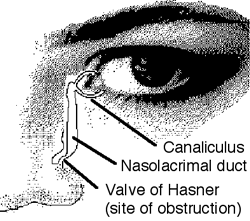
Fig
2: Rate of Spontanneous Recovery from Congential Nasolacrimal Duct Obstruction
(NLDO) without surgery: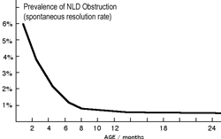
Figure
3: Anatomy of a left tear duct obstruction: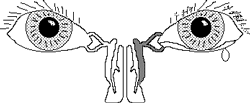
Figure
4: The "Farson" Modification of the Crawford Tube: