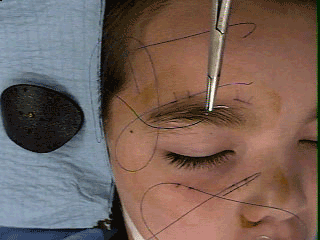
Surgical Technique for Shield Occluder
Sewing on a black shield

Scars after removal of Shield Occluder

Surgical Technique for Shield Occluder |
||
| The patient is scheduled for an "exam under anesthesia" and undergoes general anesthesia. The anatomy of the retina is carefully examined to assure potential for improvment of amblyopia in the worse eye. The properly sized shield occluder is shaped with heat to form to the orbital rim. Locations of the four pairs of suture holes are marked. The periocular skin is surgically prepared (not with Hibiclens). The 3-0 Prolene sutures are passed deep through skin but above orbicularis and periosteum and then passed through the holes in the shield occluder. Several square knots are tied and the ends of the sutures cut. Then the patient is allowed to emerge from anesthesia. | Sewing on a black shield |
|
 |
||
Scars after removal of Shield Occluder |
||
 |
||
| Parents and physicians were satisfied with clear resolution of scars after removal of the 3-0 Prolene sutures. | ||
back to Shield Occluder